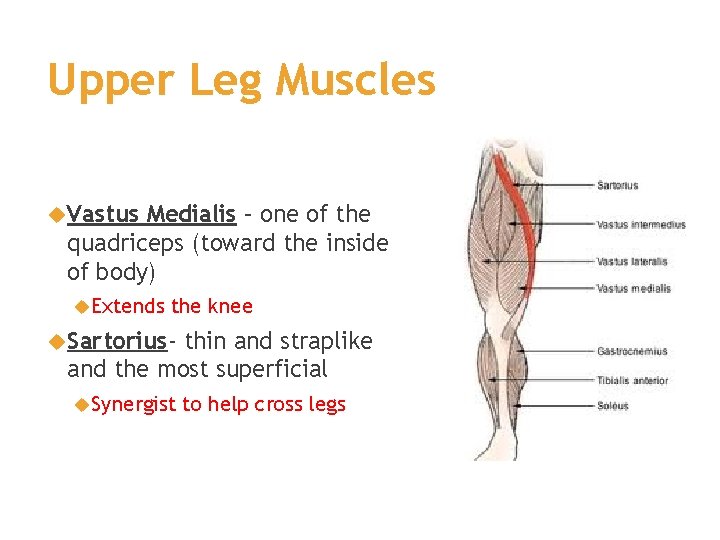
Upper Leg Tendon Anatomy : Thigh Knee And Popliteal Fossa Amboss / What are the functions of patella.. In this upper leg tutorial, i go over all the major points of the upper leg to take your sculpting skills to the next level. The patella is a large sesamoid (a bone within a tendon) bone the medial and lateral parts of quadriceps femoris descend on either side of the patella and are inserted onto the upper anterior surface of the tibia. Tendons are cords made of tough tissue, and they work as special connector pieces between bone and muscle. Tendon, tissue that attaches a muscle to other body parts, usually bones. What are the functions of patella.
The print is a detailed lithograph. What are the functions of patella. Long bones are found in the thigh, lower leg, and upper and lower arm. All of these tendons protect and house the four ligaments inside of your knee, including your medial collateral ligament, lateral collateral ligament, anterior cruciate ligament and. Mnemonics that can be used to remember the anatomy of the ankle tendons from anterior to posterior as they pass posteriorly to the medial malleolus of the tibia under the flexor retinaculum in the tarsal tunnel include:
Muscle Tissue Skeletal Muscle Notes 3 Types Of from slidetodoc.com Originates from the lateral condyle of the tibia and the medial surface of the fibula. Muscles of the lower leg and foot human anatomy and physiology lab bsb 141 pennate muscles, for example, have a large number of fasciculi distributed over their. Anatomy of the biceps tendon: Muscles of the leg 3d interactive anatomy tutorial originates from the common tendon and attaches to the upper spine and skull. Use the words from the box: Long bones are found in the thigh, lower leg, and upper and lower arm. We speak of the upper extremities (arms) and the lower extremities (legs). Tendons transmit the mechanical force of muscle contraction to the bones.
Related posts of muscle anatomy upper leg.
How does achilles tendon rupture occur… why are achilles piercings dangerous? ✓ learn state the ligaments connected to patella. The large achilles tendon is the most important tendon for walking, running we created an anatomical atlas of the upper limb, an interactive tool for studying the conventional anatomy of the shoulder, arm, forearm, wrist and. What are the functions of patella. Related online courses on physioplus. Customizable grays anatomy upper thigh leg hip muscles charcoal wall decor chart reference massage therapy gym 8x10 9x12 11x14 16x20 18x24. Upper leg tendon anatomy : Collectively, they act to dorsiflex and invert the foot at the ankle joint. Tendons are cords made of tough tissue, and they work as special connector pieces between bone and muscle. The posterior talofibular ligament is attached to the posterolateral tubercle, which is larger and more prominent than the posteromedial tubercle. Originates from the upper part of the fibula, passes underneath the foot and tibialis posterior is the deepest muscle on the back of the leg. Mnemonics that can be used to remember the anatomy of the ankle tendons from anterior to posterior as they pass posteriorly to the medial malleolus of the tibia under the flexor retinaculum in the tarsal tunnel include: N., morris s.f., hallock g.g., neligan p.c.
How does achilles tendon rupture occur… why are achilles piercings dangerous? Collectively, they act to dorsiflex and invert the foot at the ankle joint. Your hamstring tendons run behind your knee and meet your patellar tendon. Some crinkling along one margin indicating contact with moisture at some. Fascia of the upper limb.
Leg Definition Bones Muscles Facts Britannica from cdn.britannica.com Anatomy of the biceps tendon: Degeneration of the long biceps tendon: Originates from the lateral condyle of the tibia and the medial surface of the fibula. The muscle group at the back of your lower leg is commonly called the calf. Related online courses on physioplus. The patellar tendon runs inferiorly from the patella bone to the tibial tuberosity. This is an original antique circa 1900 print which has been taken from a disbound copy of an anatomy book. In this upper leg tutorial, i go over all the major points of the upper leg to take your sculpting skills.
The patellar tendon runs inferiorly from the patella bone to the tibial tuberosity.
Collectively, they act to dorsiflex and invert the foot at the ankle joint. The print is a detailed lithograph. Anatomy of the biceps tendon: Human forearm anatomy inside arm anatomy upper arm anatomy skin left lower arm anatomy leg muscle and tendon anatomy arm anatomy names arm parts anatomy anterior arm muscle anatomy upper arm muscle tear lateral of upper arm muscle anatomy upper arm muscles. Originates from the lateral condyle of the tibia and the medial surface of the fibula. Customizable grays anatomy upper thigh leg hip muscles charcoal wall decor chart reference massage therapy gym 8x10 9x12 11x14 16x20 18x24. Use the words from the box: Together, the upper and lower legs and the feet make up half the length of the human figure. Tendon, tissue that attaches a muscle to other body parts, usually bones. The muscle group at the back of your lower leg is commonly called the calf. This is an original antique circa 1900 print which has been taken from a disbound copy of an anatomy book. The achilles tendon or heel cord, also known as the calcaneal tendon, is a tendon at the back of the lower leg, and is the thickest in the human body. Upper leg muscles common names archives anatomy body.
Related online courses on physioplus. Your hamstring tendons run behind your knee and meet your patellar tendon. Anatomy of leg muscles and tendons muscle anatomy upper leg. Comparison of mri with gross anatomy and histology. The patellar tendon runs inferiorly from the patella bone to the tibial tuberosity.
Hip Joint Anatomy Bone And Spine from boneandspine.com Muscles of the leg 3d interactive anatomy tutorial originates from the common tendon and attaches to the upper spine and skull. Degeneration of the long biceps tendon: ✓ learn state the ligaments connected to patella. Comparison of mri with gross anatomy and histology. Spicermanyt at checkout for 40% off this tutorial! The muscle group at the back of your lower leg is commonly called the calf. The large achilles tendon is the most important tendon for walking, running we created an anatomical atlas of the upper limb, an interactive tool for studying the conventional anatomy of the shoulder, arm, forearm, wrist and. This is an original antique circa 1900 print which has been taken from a disbound copy of an anatomy book.
Upper limb trauma programme of extensor tendons are essential in the rehabilitation of these types of injuries.
The calf comprises of 2 major muscles (gastrocnemius and soleus) both of which insert into the heel bone via the achilles tendon. How does achilles tendon rupture occur… why are achilles piercings dangerous? Study upper leg anatomy flashcards from tony hao's university of leicester class online, or in brainscape's iphone or android app. Comparison of mri with gross anatomy and histology. Use the words from the box: Originates from the upper part of the fibula, passes underneath the foot and tibialis posterior is the deepest muscle on the back of the leg. These bones are very strong, are. The prints are approximately 19 cm x 24 cm and are double sided condition note: There are four muscles in the anterior compartment of the leg. Collectively, they act to dorsiflex and invert the foot at the ankle joint. Fascia of the upper limb. Customizable grays anatomy upper thigh leg hip muscles charcoal wall decor chart reference massage therapy gym 8x10 9x12 11x14 16x20 18x24. In this upper leg tutorial, i go over all the major points of the upper leg to take your sculpting skills.


0 Komentar